RESEARCH DIRECTION 02
Mechanical signaling and subcellular organization
We use super-resolution microscopy to understand how mechanical signaling is encoded in cellular structure. Mechanical signaling must ultimately drive physical changes in cell shape and adhesion which require nanoscale changes in cytoskeletal and membrane architecture that typically lie beyond the resolution limit of conventional light microscopy. We use an array of super-resolution microscopy and computational methods in intact cells and embryos to uncover force-induced changes in cellular architecture.
Research snapshots
Super-resolution microscopy reveals cytoskeletal architecture in intact cells and tissues
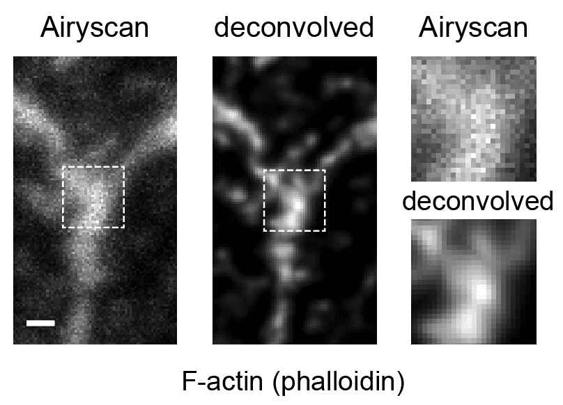
AiryScan imaging and deconvolution can reveal finer actin substructures at single tricellular junctions in developing embryos. Scale bar, 500 nm.
Taneja et al., 2026. Developmental Cell

Structured illumination microscopy reveals a population of minute actin fibers that connect nascent myofibrils to the substrate in cardiomyocytes and necessary for the formation of cardiac sarcomeres.
Taneja, Neininger, and Burnette. 2020 Molecular biology of the cell

Structured Illumination microscopy reveals altered distribution of hetero-filaments of myosin-II paralogs at the mitotic cortex of dividing cancer cells. Scale bar, 1 µm.
Taneja et al., 2020. Cell Reports